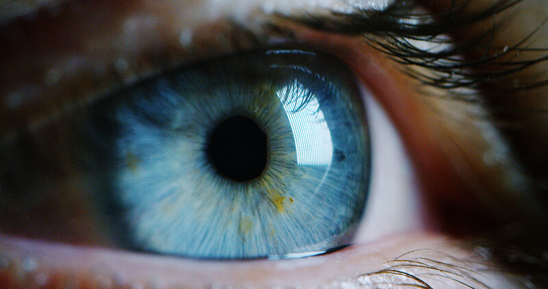
The cornea is made up of five separate layers, each with its own important function:
Epithelium
This is the cornea’s outermost layer. Its primary function is to protect the inner layers from bacteria, dust, and water. It also provides a smooth surface to absorb oxygen and nutrients from tears which are then distributed to the inner layers of the cornea.
Bowman’s Membrane
The Bowman’s membrane is an extremely thin, clear film of tissue made of strong protein fibers called collagen. When injured, the Bowman’s layer has a high risk of scarring which can lead to vision loss.
Stroma
Behind the Bowman’s layer lies the stroma. The stroma is the thickest layer of the cornea, making up about 90% of the corneal thickness. The stroma is composed of water and collagen which is important in maintaining its transparency and shape.
Descemet’s Membrane
Behind the stroma is the Descemet’s membrane. This thin, strong membrane protects against infection and injury and repairs itself easily after injury.
Endothelium
The endothelium is the innermost layer. This thin layer contains endothelial cells which are important in keeping the cornea clear. Fluid from inside the eye leaks slowly into the stroma. The endothelium is responsible for pumping excess fluid out of the stroma. Without a healthy endothelium, fluid would build-up, leading to swelling of the cornea and causing vision loss (a condition called Fuch’s dystrophy).

Common Cornea Diseases and Disorders
The cornea, while resilient, is also susceptible to many different diseases and disorders:
- Allergies
- Injuries
- Keratitis
- Dry Eye Syndrome
- Dystrophies
- Keratoconus
- Fuch’s Dystrophy
- Pterygium
Corneal Cross Linking
The team at Pacific Eye Institute is now performing cross-linking procedures to strengthen and stabilize the shape of your cornea. The procedure aims to stop the cornea from getting thinner, weaker, and more irregular in shape. It is highly effective in slowing disease progression and stabilizing existing vision.
This is an in-office procedure that treats both keratoconus and post-LASIK corneal ectasia. In a short, painless procedure, our surgeon will work to increases the rigidness of the cornea’s surface by inducing additional cross-links between collagen fibers.
We gently remove the cornea’s epithelial layer and then applies riboflavin (B2) eye drops to the surface of the eye. Controlled ultraviolet light is then used to treat the eye. After the treatment, your eye doctor will apply a special contact lens to protect the eye and prescribe antibiotic and anti-inflammatory eye drops.
Corneal Transplant
If the cornea becomes damaged beyond repair, a corneal transplant may be necessary. This process works by removing all or part of the cornea and replacing it with healthy donor tissue from an eye bank. The corneal specialists at Pacific Eye Institute can assess the health of your cornea and make treatment recommendations:
- Full-thickness corneal transplant (penetrating keratoplasty or PK) to replace the entire cornea
- Partial thickness corneal transplant (Descemet’s Stripping Automated Endothelial Keratoplasty or DSAEK) which replaces the damaged section of the back inner layer of the cornea
- Descemet Membrane Endothelial Keratoplasty (DMEK) is a partial-thickness cornea transplant procedure with selective removal of the patient’s Descemet membrane and endothelium, followed by transplantation of donor corneal tissue. DMEK as an advanced corneal transplant procedure, offering faster recovery times and better visual outcomes.
Tears and Your Cornea
Each time we blink, a thin layer of tears spreads over the eye. This tear film allows our eyelids to move smoothly over the corneal surface as well as prevents infection and promotes healing. The tear film consists of three components: an outer oily (lipid) layer, a middle water (aqueous) layer, and a bottom mucin layer. Each component of the tear film is important in overall eye health:
- Lipid layer. The outermost oil (lipid) layer keeps tears from evaporating too quickly from the eye. When the oil layer is lacking, it leads to a condition called evaporative dry eye syndrome.
- Aqueous layer. The middle aqueous layer nourishes the cornea and conjunctiva.
- Mucin layer. The bottom mucin layer helps spread the aqueous layer over the eye, ensuring that the eye remains wet.
When a component of the tear film is not adequate, it can result in irritation and redness. Dry eye syndrome may also develop. The tear film is extremely important to eye health, so any prolonged dryness or redness of your eyes must be reported to your eye doctor.
Dry Eye Syndrome
Dry eye syndrome is extremely common. It occurs when the eye either cannot produce enough tears or produces poor-quality tears. Symptoms of dry eye syndrome generally include a feeling of dryness, redness, the feeling that something is in your eye, and pain in the eye. Mild to moderate dry eye symptoms can usually be managed with over-the-counter lubricating eye drops. Severe cases of dry eye syndrome should be treated by a doctor because prolonged dryness can cause more serious problems over a long period. To learn more about our dry eye treatment options and Lipiflow, visit our Dry Eye Pages
To learn more about cornea treatments in the Inland Empire, contact Pacific Eye Institute today to schedule an eye exam.


